|
|
Brain StudiesBrain Basics:Know Your Brain & The Life and Death of a Neuron
|
A continuing education course for 1 ce
Brain
Basics
Know
Your Brain & The
Life and Death of a Neuron
Brain
Basics: Know Your Brain
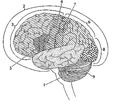
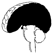
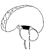
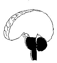
IntroductionThe brain is the most complex part of the human body. This three-pound organ is the seat of intelligence, interpreter of the senses, initiator of body movement, and controller of behavior. Lying in its bony shell and washed by protective fluid, the brain is the source of all the qualities that define our humanity. The brain is the crown jewel of the human body. For centuries, scientists and philosophers have been fascinated by the brain, but until recently they viewed the brain as nearly incomprehensible. Now, however, the brain is beginning to relinquish its secrets. Scientists have learned more about the brain in the last 10 years than in all previous centuries because of the accelerating pace of research in neurological and behavioral science and the development of new research techniques. As a result, Congress named the 1990s the Decade of the Brain. At the forefront of research on the brain and other elements of the nervous system is the National Institute of Neurological Disorders and Stroke (NINDS), which conducts and supports scientific studies in the United States and around the world. This fact sheet is a basic introduction to the human brain. It may help you understand how the healthy brain works, how to keep it healthy, and what happens when the brain is diseased or dysfunctional. <top> The Architecture of the Brain The brain is like a committee of experts. All the parts of the brain work together, but each part has its own special properties. The brain can be divided into three basic units: the forebrain, the midbrain, and the hindbrain. The hindbrain includes the upper part of the spinal cord, the brain stem, and a wrinkled ball of tissue called the cerebellum (1). The hindbrain controls the body’s vital functions such as respiration and heart rate. The cerebellum coordinates movement and is involved in learned rote movements. When you play the piano or hit a tennis ball you are activating the cerebellum. The uppermost part of the brainstem is the midbrain, which controls some reflex actions and is part of the circuit involved in the control of eye movements and other voluntary movements. The forebrain is the largest and most highly developed part of the human brain: it consists primarily of the cerebrum (2) and the structures hidden beneath it (see "The Inner Brain"). When people see pictures of the brain it is usually the cerebrum that they notice. The cerebrum sits at the topmost part of the brain and is the source of intellectual activities. It holds your memories, allows you to plan, enables you to imagine and think. It allows you to recognize friends, read books, and play games. The cerebrum is split into two halves (hemispheres) by a deep fissure. Despite the split, the two cerebral hemispheres communicate with each other through a thick tract of nerve fibers that lies at the base of this fissure. Although the two hemispheres seem to be mirror images of each other, they are different. For instance, the ability to form words seems to lie primarily in the left hemisphere, while the right hemisphere seems to control many abstract reasoning skills. For some as-yet-unknown reason, nearly all of the signals from the brain to the body and vice-versa cross over on their way to and from the brain. This means that the right cerebral hemisphere primarily controls the left side of the body and the left hemisphere primarily controls the right side. When one side of the brain is damaged, the opposite side of the body is affected. For example, a stroke in the right hemisphere of the brain can leave the left arm and leg paralyzed. The Forebrain ------- The Midbrain -------- The Hindbrain
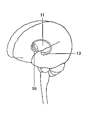
<top> The Geography of Thought Each cerebral hemisphere can be divided into sections, or lobes, each of which specializes in different functions. To understand each lobe and its specialty we will take a tour of the cerebral hemispheres, starting with the two frontal lobes (3), which lie directly behind the forehead. When you plan a schedule, imagine the future, or use reasoned arguments, these two lobes do much of the work. One of the ways the frontal lobes seem to do these things is by acting as short-term storage sites, allowing one idea to be kept in mind while other ideas are considered. In the rearmost portion of each frontal lobe is a motor area (4), which helps control voluntary movement. A nearby place on the left frontal lobe called Broca’s area (5) allows thoughts to be transformed into words. When you enjoy a good meal—the taste, aroma, and texture of the food—two sections behind the frontal lobes called the parietal lobes (6) are at work. The forward parts of these lobes, just behind the motor areas, are the primary sensory areas (7). These areas receive information about temperature, taste, touch, and movement from the rest of the body. Reading and arithmetic are also functions in the repertoire of each parietal lobe. As you look at the words and pictures on this page, two areas at the back of the brain are at work. These lobes, called the occipital lobes (8), process images from the eyes and link that information with images stored in memory. Damage to the occipital lobes can cause blindness. The last lobes on our tour of the cerebral hemispheres are the temporal lobes (9), which lie in front of the visual areas and nest under the parietal and frontal lobes. Whether you appreciate symphonies or rock music, your brain responds through the activity of these lobes. At the top of each temporal lobe is an area responsible for receiving information from the ears. The underside of each temporal lobe plays a crucial role in forming and retrieving memories, including those associated with music. Other parts of this lobe seem to integrate memories and sensations of taste, sound, sight, and touch. <top> The Cerebral Cortex Coating the surface of the cerebrum and the cerebellum is a vital layer of tissue the thickness of a stack of two or three dimes. It is called the cortex, from the Latin word for bark. Most of the actual information processing in the brain takes place in the cerebral cortex. When people talk about "gray matter" in the brain they are talking about this thin rind. The cortex is gray because nerves in this area lack the insulation that makes most other parts of the brain appear to be white. The folds in the brain add to its surface area and therefore increase the amount of gray matter and the quantity of information that can be processed. <top> The Inner Brain Deep within the brain, hidden from view, lie structures that are the gatekeepers between the spinal cord and the cerebral hemispheres. These structures not only determine our emotional state, they also modify our perceptions and responses depending on that state, and allow us to initiate movements that you make without thinking about them. Like the lobes in the cerebral hemispheres, the structures described below come in pairs: each is duplicated in the opposite half of the brain. The hypothalamus (10), about the size of a pearl, directs a multitude of important functions. It wakes you up in the morning, and gets the adrenaline flowing during a test or job interview. The hypothalamus is also an important emotional center, controlling the molecules that make you feel exhilarated, angry, or unhappy. Near the hypothalamus lies the thalamus (11), a major clearinghouse for information going to and from the spinal cord and the cerebrum. An arching tract of nerve cells leads from the hypothalamus and the thalamus to the hippocampus (12). This tiny nub acts as a memory indexer—sending memories out to the appropriate part of the cerebral hemisphere for long-term storage and retrieving them when necessary. The basal ganglia (not shown) are clusters of nerve cells surrounding the thalamus. They are responsible for initiating and integrating movements. Parkinson’s disease, which results in tremors, rigidity, and a stiff, shuffling walk, is a disease of nerve cells that lead into the basal ganglia.
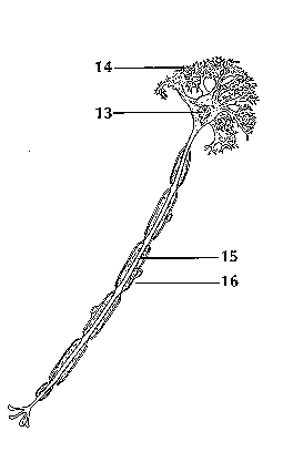
<top> Making Connections The brain and the rest of the nervous system are composed of many different types of cells, but the primary functional unit is a cell called the neuron. All sensations, movements, thoughts, memories, and feelings are the result of signals that pass through neurons. Neurons consist of three parts. The cell body (13) contains the nucleus, where most of the molecules that the neuron needs to survive and function are manufactured. Dendrites (14) extend out from the cell body like the branches of a tree and receive messages from other nerve cells. Signals then pass from the dendrites through the cell body and may travel away from the cell body down an axon (15) to another neuron, a muscle cell, or cells in some other organ. The neuron is usually surrounded by many support cells. Some types of cells wrap around the axon to form an insulating sheath (16). This sheath can include a fatty molecule called myelin, which provides insulation for the axon and helps nerve signals travel faster and farther. Axons may be very short, such as those that carry signals from one cell in the cortex to another cell less than a hair’s width away. Or axons may be very long, such as those that carry messages from the brain all the way down the spinal cord.
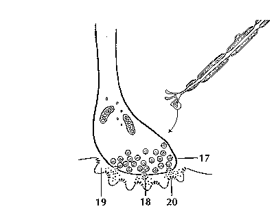
Scientists have learned a great deal about neurons by studying the synapse—the place where a signal passes from the neuron to another cell. When the signal reaches the end of the axon it stimulates tiny sacs (17). These sacs release chemicals known as neurotransmitters (18) into the synapse (19). The neurotransmitters cross the synapse and attach to receptors (20) on the neighboring cell. These receptors can change the properties of the receiving cell. If the receiving cell is also a neuron, the signal can continue the transmission to the next cell.
<top> Some Key Neurotransmitters at Work Acetylcholine is called an excitatory neurotransmitter because it generally makes cells more excitable. It governs muscle contractions and causes glands to secrete hormones. Alzheimer’s disease, which initially affects memory formation, is associated with a shortage of acetylcholine. GABA (gamma-aminobutyric acid) is called an inhibitory neurotransmitter because it tends to make cells less excitable. It helps control muscle activity and is an important part of the visual system. Drugs that increase GABA levels in the brain are used to treat epileptic seizures and tremors in patients with Huntington’s disease. Serotonin is an inhibitory neurotransmitter that constricts blood vessels and brings on sleep. It is also involved in temperature regulation. Dopamine is an inhibitory neurotransmitter involved in mood and the control of complex movements. The loss of dopamine activity in some portions of the brain leads to the muscular rigidity of Parkinson’s disease. Many medications used to treat behavioral disorders work by modifying the action of dopamine in the brain. <top> Neurological Disorders When the brain is healthy it functions quickly and automatically. But when problems occur, the results can be devastating. Some 50 million people in this country—one in five—suffer from damage to the nervous system. The NINDS supports research on more than 600 neurological diseases. Some of the major types of disorders include: neurogenetic diseases (such as Huntington’s disease and muscular dystrophy), developmental disorders (such as cerebral palsy), degenerative diseases of adult life (such as Parkinson’s disease and Alzheimer’s disease), metabolic diseases (such as Gaucher’s disease), cerebrovascular diseases (such as stroke and vascular dementia), trauma (such as spinal cord and head injury), convulsive disorders (such as epilepsy), infectious diseases (such as AIDS dementia), and brain tumors. <top> The National Institute of Neurological Disorders and Stroke Since its creation by Congress in 1950, the NINDS has grown to become the leading supporter of neurological research in the United States. Most research funded by the NINDS is conducted by scientists in public and private institutions such as universities, medical schools, and hospitals. Government scientists also conduct a wide array of neurological research in the more than 20 laboratories and branches of the NINDS itself. This research ranges from studies on the structure and function of single brain cells to tests of new diagnostic tools and treatments for those with neurological disorders. For information on other neurological disorders or research programs funded by the National Institute of Neurological Disorders and Stroke, contact the Institute's Brain Resources and Information Network (BRAIN) at: BRAIN
NIH Publication No.01-3440a Last updated May 01, 2007 |

The
Life and Death of a NeuronTable of Contents
IntroductionUntil recently, most neuroscientists thought we were born with all the neurons we were ever going to have. As children we might produce some new neurons to help build the pathways - called neural circuits - that act as information highways between different areas of the brain. But scientists believed that once a neural circuit was in place, adding any new neurons would disrupt the flow of information and disable the brain’s communication system. In 1962, scientist Joseph Altman challenged this belief when he saw evidence of neurogenesis (the birth of neurons) in a region of the adult rat brain called the hippocampus. He later reported that newborn neurons migrated from their birthplace in the hippocampus to other parts of the brain. In 1979, another scientist, Michael Kaplan, confirmed Altman’s findings in the rat brain, and in 1983 he found neural precursor cells in the forebrain of an adult monkey. These discoveries about neurogenesis in the adult brain were surprising to other researchers who didn’t think they could be true in humans. But in the early 1980s, a scientist trying to understand how birds learn to sing suggested that neuroscientists look again at neurogenesis in the adult brain and begin to see how it might make sense. In a series of experiments, Fernando Nottebohm and his research team showed that the numbers of neurons in the forebrains of male canaries dramatically increased during the mating season. This was the same time in which the birds had to learn new songs to attract females. Why did these bird brains add neurons at such a critical time in learning? Nottebohm believed it was because fresh neurons helped store new song patterns within the neural circuits of the forebrain, the area of the brain that controls complex behaviors. These new neurons made learning possible. If birds made new neurons to help them remember and learn, Nottebohm thought the brains of mammals might too. Other scientists believed these findings could not apply to mammals, but Elizabeth Gould later found evidence of newborn neurons in a distinct area of the brain in monkeys, and Fred Gage and Peter Eriksson showed that the adult human brain produced new neurons in a similar area. For some neuroscientists, neurogenesis in the adult brain is still an unproven theory. But others think the evidence offers intriguing possibilities about the role of adult-generated neurons in learning and memory.
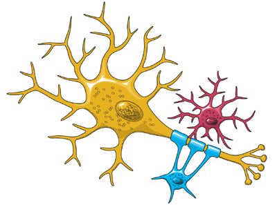
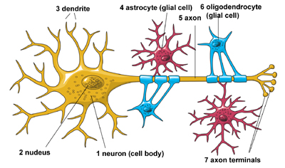
The Architecture of the NeuronThe central nervous system (which includes the brain and spinal cord) is made up of two basic types of cells: neurons (1) and glia (4) & (6). Glia outnumber neurons by a substantial amount -- some scientists have estimated it to be as large as nine to one -- but in spite of their smaller numbers, neurons are the key players in the brain. Neurons are information messengers. They use electrical impulses and chemical signals to transmit information between different areas of the brain, and between the brain and the rest of the nervous system. Everything we think and feel and do would be impossible without the work of neurons and their support cells, the glial cells called astrocytes (4) and oligodendrocytes (6). Neurons have three basic parts: a cell body and two extensions called an axon (5) and a dendrite (3). Within the cell body is a nucleus (2), which controls the cell’s activities and contains the cell’s genetic material. The axon looks like a long tail and transmits messages from the cell. Dendrites look like the branches of a tree and receive messages for the cell. Neurons communicate with each other by sending chemicals, called neurotransmitters, across a tiny space, called a synapse, between the axons and dendrites of adjacent neurons.
There are three classes of neurons:
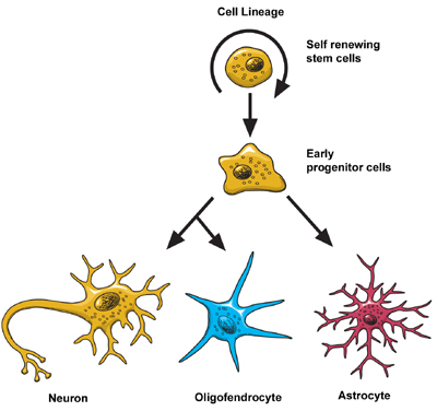
Scientists think that neurons are the most diverse kind of cell in the body. Within these three classes of neurons are hundreds of different types, each with specific message-carrying abilities. How these neurons communicate with each other by making connections is what makes each of us unique in how we think, and feel, and act. BirthThe extent to which new neurons are generated in the brain is a controversial subject among neuroscientists. Although the majority of neurons are already present in our brains by the time we are born, there is evidence to support that neurogenesis (the scientific word for the birth of neurons) is a lifelong process. Neurons are born in areas of the brain that are rich in concentrations of neural precursor cells (also called neural stem cells). These cells have the potential to generate most, if not all, of the different types of neurons and glia found in the brain. Neuroscientists have observed how neural precursor cells behave in the laboratory. Although this may not be exactly how these cells behave when they are in the brain, it gives us information about how they could be behaving when they are in the brain’s environment. The science of stem cells is still very new, and could change with additional discoveries, but researchers have learned enough to be able to describe how neural stem cells generate the other cells of the brain. They call it a stem cell’s lineage and it is similar in principle to a family tree. Neural stem cells increase by dividing in two and producing either two new stem cells, or two early progenitor cells, or one of each. When a stem cell divides to produce another stem cell, it is said to self-renew. This new cell has the potential to make more stem cells. When a stem cell divides to produce an early progenitor cell, it is said to differentiate. Differentiation means that the new cell is more specialized in form and function. An early progenitor cell does not have the potential of a stem cell to make many different types of cells. It can only make cells in its particular lineage. Early progenitor cells can self-renew or go in either of two ways. One type will give rise to astrocytes. The other type will ultimately produce neurons or oligodendrocytes. MigrationOnce a neuron is born it has to travel to the place in the brain where it will do its work. How does a neuron know where to go? What helps it get there? Scientists have seen that neurons use at least two different methods to travel:
Not all neurons are successful in their journey. Scientists think that only a third reach their destination. The rest either never differentiate, or die and disappear at some point during the two to three week phase of migration. Some neurons survive the trip, but end up where they shouldn’t be. Mutations in the genes that control migration create areas of misplaced or oddly formed neurons that can cause disorders such as childhood epilepsy or mental retardation. Some researchers suspect that schizophrenia and the learning disorder dyslexia are partly the result of misguided neurons.
DifferentiationOnce a neuron reaches its destination, it has to settle in to work. This final step of differentiation is the least well-understood part of neurogenesis. Neurons are responsible for the transport and uptake of neurotransmitters - chemicals that relay information between brain cells. Depending on its location, a neuron can perform the job of a sensory neuron, a motor neuron, or an interneuron, sending and receiving specific neurotransmitters. In the developing brain, a neuron depends on molecular signals from other cells, such as astrocytes, to determine its shape and location, the kind of transmitter it produces, and to which other neurons it will connect. These freshly born cells establish neural circuits - or information pathways connecting neuron to neuron - that will be in place throughout adulthood. But in the adult brain, neural circuits are already developed and neurons must find a way to fit in. Researchers suspect that astrocytes play a similar role in the adult brain, actively regulating the function and synapse formation of new neurons. As a new neuron settles in, it starts to look like surrounding cells. It develops an axon and dendrites and begins to communicate with its neighbors.
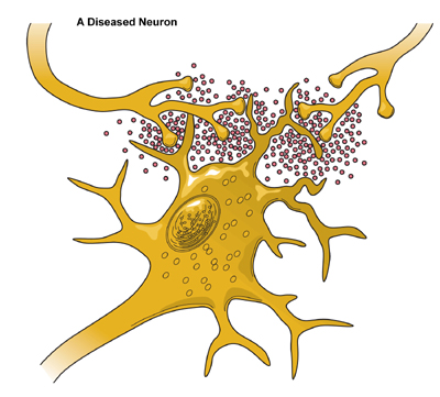
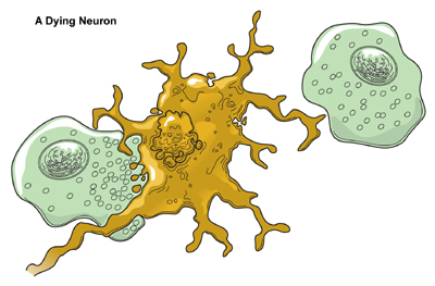
DeathAlthough neurons are the longest living cells in the body, large numbers of them die during migration and differentiation. The lives of some neurons can take abnormal turns. Some diseases of the brain are the result of the unnatural deaths of neurons. - In Parkinson’s disease, neurons that produce the neurotransmitter dopamine die off in the basal ganglia, an area of the brain that controls body movements. The brain can no longer control the body and people shake and jerk in spasms. - In Huntington’s disease, a genetic mutation causes over-production of a neurotransmitter called glutamate, which kills neurons in the basal ganglia. As a result, people twist and writhe uncontrollably. - In Alzheimer’s disease, unusual proteins build up in and around neurons in the neocortex and hippocampus, parts of the brain that control memory. When these neurons die, people lose their capacity to remember and their ability to do everyday tasks. Physical damage to the brain and other parts of the central nervous system can also kill or disable neurons. - Blows to the brain, or the damage caused by a stroke, can kill neurons outright or slowly starve them of the oxygen and nutrients they need to survive. - Spinal cord injury can disrupt communication between the brain and muscles when neurons lose their connection to axons located below the site of injury. These neurons may still live, but they lose their ability to communicate.
Hope Through ResearchScientists hope that by understanding more about the life and death of neurons they can develop new treatments, and possibly even cures, for brain diseases and disorders that affect the lives of millions of Americans. The most current research suggests that neural stem cells can generate many, if not all, of the different types of neurons found in the brain and the nervous system. Learning how to manipulate these stem cells in the laboratory into specific types of neurons could produce a fresh supply of brain cells to replace those that have died or been damaged. Therapies could also be created to take advantage of growth factors and other signaling mechanisms inside the brain that tell precursor cells to make new neurons. This would make it possible to repair, reshape, and renew the brain from within. For information on other neurological disorders or research programs funded by the National Institute of Neurological Disorders and Stroke, contact the Institute's Brain Resources and Information Network (BRAIN) at: BRAIN Prepared
by: NINDS health-related material is provided for information purposes only and does not necessarily represent endorsement by or an official position of the National Institute of Neurological Disorders and Stroke or any other Federal agency. Advice on the treatment or care of an individual patient should be obtained through consultation with a physician who has examined that patient or is familiar with that patient's medical history. All NINDS-prepared information is in the public domain and may be freely copied. Credit to the NINDS or the NIH is appreciated. |
||||||||||||||||||||||||||||||||||||
maintains responsibility for the program. |
Resources: |
We do adhere to the American Psychological Association's Ethical Principles of Psychologists. Our courses are carefully screened by the Planning Committee to adhere to APA standards. We also require authors who compose Internet courses specifically for us follow APA ethical standards. Many of our courses contain case material, and may use the methods of qualitative research and analysis, in-depth interviews and ethnographic studies. The psychotherapeutic techniques depicted may include play therapy, sandplay therapy, dream analysis, drawing analysis, client and therapist self-report, etc. The materials presented may be considered non-traditional and may be controversial, and may not have widespread endorsement within the profession. www.psychceu.com maintains responsibility for the program and its content. |
All material included in this course is either in the public domain, or used with express permission. |
![]()
|
Thank you! |
To
take the post-test, please return to
http://www.psychceu.com/materialsandtests/login.asp
888-777-3773
|
|
|
|